Hexamita and Spironucleus
جهت مشاهده متن کامل مقاله اینجا کلیک کنید
Hexamita and Spironucleus
- reported in the digestive tract of fishes(sometimes in internal organs)
- direct life cycles
- they are difficult to distinguish under the light microscope
- known as hexamitosis, spironucleosis and possibly ‘hole-in-the head’ disease in salmonids, cyprinids and ornamental aquarium fishes
- Sporadic outbreaks of fatal systemic spironucleosis have occurred in cyprinids ,eels ,tropical aquarium fishes, Atlantic salmon and chinook salmon
Geographical distribution and host range:
- very wide host range and geographical distribution
- These parasites are in wild, farmed and aquarium fishes in cold, temperate and warm waters.
- They have been reported in Acipenseridae, Anguillidae, Catostomidae, Centarchidae, Cyprinidae, Cyprinodontidae, Gadidae, Gasterosteidae, Mugilidae, Percichthyidae, Percidae, Salmonidae, Siganidae and Sparidae
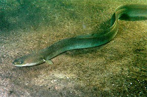
- Hexamita salmonis has been reported in fishes in North America, Europe and Asia. It is usually found in freshwater fishes but it has also been reported from marine fishes.
–The disease is most commonly found in fingerlings, although yearlings and smolts may also show clinical signs of the infection.
–Outbreaks of the disease are usually sporadic in aquaculture facilities, and they have seldom been reported in wild fishes
- Spironucleus vortens is in angelfish Florida, in discus and in and in ide (Leuciscus idus).
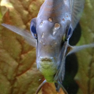
–causative agent in ‘hole-in-the-head’ disease in cichlids. The parasite is also known as ‘discus parasite’ as it is often found in discus fish
- Spironucleus are known to be pathogenic and cause disease in salmonids maintained in sea cages. Spironucleus barkhanus has been isolated from Atlantic salmon (Salmo salar), grayling (Thymallus thymallus) and Arctic charr (Salvelinus alpinus) in Norway
Morphology:
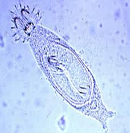
- Live trophozoites of Hexamita and Spironucleus vary from elongated to nearly spherical and the two genera are usually difficult to separate.
- The lengths of live organisms may reach 20 μm and the organisms have bilateral symmetry
- Hexamita has an elongated to spherical body and spherical nuclei, while a pyriform body and elongated nuclei are characteristic of Spironucleus
Diagram of a typical diplomonad flagellate with diagnostic features: size (from 5 to 20 μ m long, excluding the flagella); eight flagella (three pairs anteriorly, one pair posteriorly); pyriform to ellipsoidal to egg - shape to tapering body.
- the two recurrent flagella in their canals are located between the nuclei in Spironucleus while those in Hexamita are on the surface of the nuclei
- Diplomonad cysts are usually oval in shape, and each cyst usually contains two recently divided flagellates.
Scanning electron micrographs of Spironucleus vortens. Normal, untreated trophozoites are pyriform-shaped possessing 8 flagella
Clinical signs and pathology:
- Intestinal infections: Trophozoites are commonly found in the lumen of the intestine, while cysts are often not seen.
- the flagellate interfered with nutrition by competing for essential nutrients and/or by damaging the intestinal epithelium to affect digestion and absorption.
- Systemic hexamitid infections have been reported in cyprinids, eels, salmonids and tropical aquarium freshwater fishes , with trophozoites in the blood, gall bladder, heart, kidney, liver, spleen, eye, brain, muscles, mesentery and abdominal cavity. There are also reports of hexamitids in cranial skeletal tissues and in pustules on the skin of infected cichlids
- weakness, whirling, listlessness, abdominal distension, anaemia, faecal pseudocasts, pale stringy faeces, a red vent, exophthalmia and dark coloration
- Fish infected with H. salmonis may have anaemia, ascites, enteritis and intestinal contents that are yellowish and watery or jelly-like, with excessive amounts of mucus.
- Infected aquarium tropical fish may have yellow mucus in their intestines, enteritis and enlarged and inflamed gall bladder.
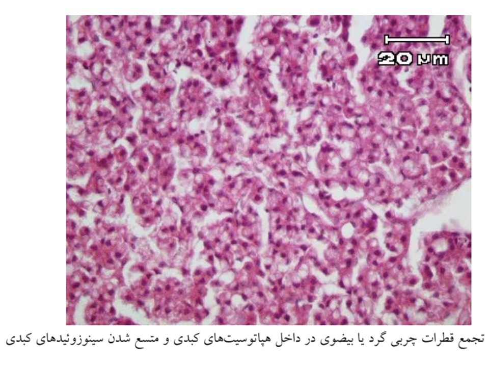
- The pathology associated with the infection varies from no effects to haemorrhages in the intestine , catarrhal enteritis and hepatocellular necrosis
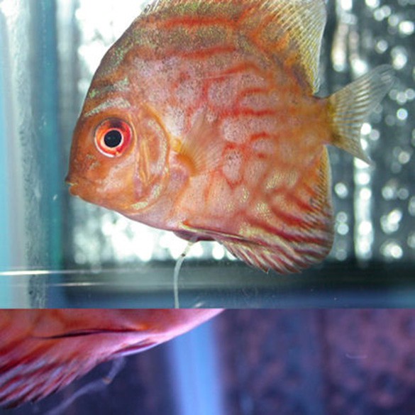
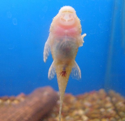
This Tiger Oscar has succumbed to hole in the head disease
Oscar with hole in the head disease
Diagnosis
- Wet mount of skin, feces, or viscera with parasites
- Histopathology of lesion with parasites
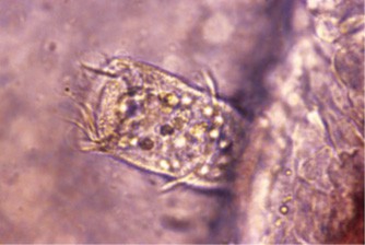
Wet mount of diplomonds
Figs. 10 & 11. Salvelinus alpinus. Scanning electron micrographs of blood from Arctic char showing presence of trophozoites of Spironucleus barkhanus. Scale bar in 10 = 10 μm;in 11 = 2 μm