Viral hemorrhagic septicemia
جهت مشاهده متن کامل مقاله اینجا کلیک کنید
Viral hemorrhagic septicemia (vhs; egteved disease)
Introduction
VHSV is a bullet shaped particle measuring approximately 180nm by 70nm. It is surrounded by a noticeable envelope and this contains the viral membrane glycoprotein, which is the main surface antigen involved in neutralization by antibody. The virion RNA is a single-stranded molecule of negative sense polarity
Viral hemorrhagic septicemia, caused by a rhabdovirus of the genus Novirhabdovirus, is a major cause of mortality in salmonids in freshwater
The disease was first described in rainbow trout by schaperclaus (1938)
It is primarily a disease of rainbow trout and brown trout. Atlantic salmon, brook trout, and golden trout are experimentally susceptible
Previously confined to Europe, VHSV has since been isolated from asymptomatic steelhead trout, coho and chinook salmon in Puget Sound and the Gulf of Alaska, United States
In addition; VHSV has been isolated from an increasingly large number of other fish that currently includes over 50 species (mostly marine) in North America, Asia and Europe
During VHS epidemics, any age salmonid can become clinically ill, but young fish are most severely affected. Mortality can be up to 100% in fry and often ∼30 – 70% in older fish
Epidemics typically occur in spring when temperatures are fluctuating and generally below 14ᵒC
During epidemics, virus is readily transmitted horizontally by water; virus is shed in the urine and possibly from the gills but not from the feces
Ingestion of virus does not appear to be a route of transmission in salmonids, but rather gills and possibly skin wounds
The surface of eggs released by latent carriers can carry virus, but it is lost within hours; thus, vertical transmission has not been demonstrated
Gene probes can distinguish four genetic strains of VHSV, which appear to group together according to geographic region, rather than host species:
Genotype I: includes European VHSV freshwater isolates and a group of isolates from northern European waters (Baltic Sea);
Genotype II: includes another group of marine isolates from the Baltic Sea;
Genotype III: includes isolates from the North Sea, Skagerrak and Kattegat;
Genotype IV: includes North American isolates and isolates from Japan and Korea
There are three phases to VHS outbreaks, which reflect the severity of infection, not the chronological stages:
The Acute Phase
The Chronic Phase
The Nervous Phase
The acute phase (typically at 15– 18 °C) involves rapid, initially high mortality, but lower cumulative mortality
Fish are dark and lethargic (congregate away from the current on the edges of the pond or raceway, eventually massing near the outlet screen), with reddening at the base of the fins and gills caused by injection of vessels and punctate hemorrhage
There is also hemorrhage in the abdominal cavity and a leucopenia
In the chronic phase (typically at 1– 5 ° C), there are moderate, mainly protracted deaths that eventually result in high cumulative mortality
Fish are black, with anemia, exophthalmos, and a swollen abdomen
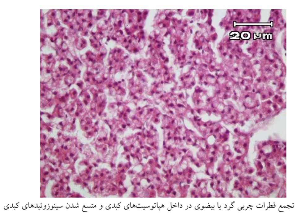
The organs are pale from severe anemia, with organizing hemorrhages. The kidney and liver may be swollen
In the nervous phase, there are low mortalities. Fish do not have gross lesions but exhibit a looping swimming behavior, darting through the water and spiraling at the bottom of the pond
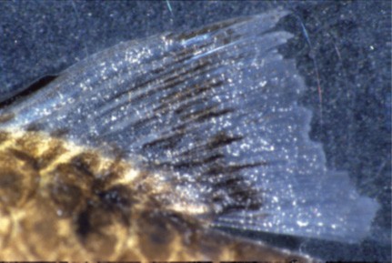
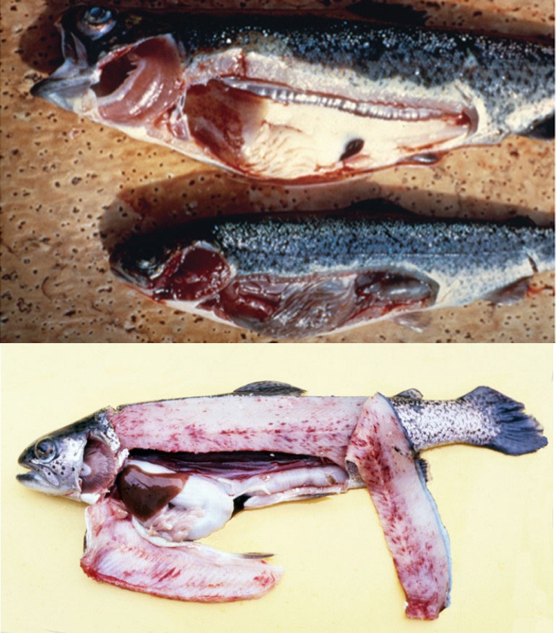
Fig 1. A. VHS in trout, showing punctate hemorrhage. B. Chronic VHS in trout (top fish), showing exophthalmos and anemia (pale gills and viscera).
(A and B photographs courtesy of the National Fish Health Research Laboratory, USA.)
Fig 2. Viral haemorrhagic septicaemia in rainbow trout showing widespread petechiae of the musculature and pale gills.