نکروز عفونی مراکز خونساز
نکروز عفونی مراکز خونساز
Infectious hematopoietic necrosis
متن کامل مقاله اینجا کلیک کنید
ویژگیهای ویروس
- رابدوویروس واجد ویریون بدون بند و یکپارچه
- ویریون دارای بار منفی
- RNA تک رشته ای
- 11000 نوکلئوتید
- 6 پروتئین رمز دار به نام های N,P,M,G,NV,L
- معروف ترین سویه آن WRAC
- جداسازی ویروس به شدت به مناطق جغرافیایی وابسته بوده و به میزان و یا سال جداسازی ویروس ارتباط ندارد
- بین ژنوتیپ ها و سروتیپ ها ارتباط کمی وجود دارد و یا حتی وجود ندارد
- حساس به حرارت، اسید، اتر، ضدعفونی کننده های رایج و خشک کردن
- اگر مواد آلی در آب باشد می تواند حداقل یک ماه در آب شیرین سرد زنده بماند. در آب شور بیشتر می تواند زنده بماند
- از طریق ادرار، مایعات جنسی و مخاط خارجی بدن دفع می شود
- در بافت های کلیه ، طحال و سایر اندام های داخلی ماهیان آلوده شده استقرار می یابد
- مرحله لاروی حساس ترین مرحله جهت ابتلا به بیماری است
- با افزایش سن مقاوم می شوند ولی باشروع تخم ریزی دوباره حساس شده و دفع ویروس افزایش می یابد
- ماهیان نجات یافته از بیماری ایمنیت بالایی در ابتلا مجدد به بیماری پیدا می کنند
- عوامل محیطی در بروز بیماری و تظاهر فرم تحت بالینی به فرم بالینی نقش مهمی دارند
- در دمای معمول آب طول مدت حامل بودن ماهی کوتاه است
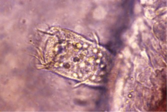
- ناقلین بی مهره مثل شپشک آبشش، زالوها و حشرات در حمل ویروس نقش دارند
- انتقال عمودی تایید نشده ولی از تخمدان جدا شده است
- مرگ به دلیل ایجاد عدم تعادل اسمزی
- تکثیر ویروس در سلول های اندوتلیال عروق، بافت های خونساز و سلول های کلیوی صورت می گیرد و باعث بروز علائم بالینی می شودعلایم عمدتا با ادم و خونریزی همراه است
- در آزاد ماهیان وحشی سواحل غربی آمریکای شمالی،IHNV بومی است
- در ایران برای اولین بار در سال 2003 جداسازی و شناسایی شد
- از نیمکره جنوبی تاکنون گزارش نشده
- تلفات بسته به شرایط محیط، درجه حرارت و میزبان در فرم حاد تا 95 درصد هم می رسد
- مهم ترین فاکتور دخیل در بروز و پیشرفت بیماری درجه حرارت آب است
- در دماهای بین 18 – 3 دیده شده ولی بطور معمول بین 15 – 8 درجه سانتیگراد دیده
- مطالعات خوبی در خصوص واکسن انجام شده ولی نیازمند بررسی های بیشتر است
- دوره کمون از 4 روز تا 2 هفته
- کست های مدفوعی در فرم مزمن از تفاوت های این بیماری با VHS است
- بهترین روش تشخیص کشت و جداسازی ویروس است
- در یک سال اخیر دوباره در ایران شیوع پیدا کرده
- ماهیان ماده نقش بیشتری در انتشار ویروس دارند
- بافت های خونساز برای کشت سلولی خیلی خوب جواب می دهند
علائم بالینی
- بی حالی
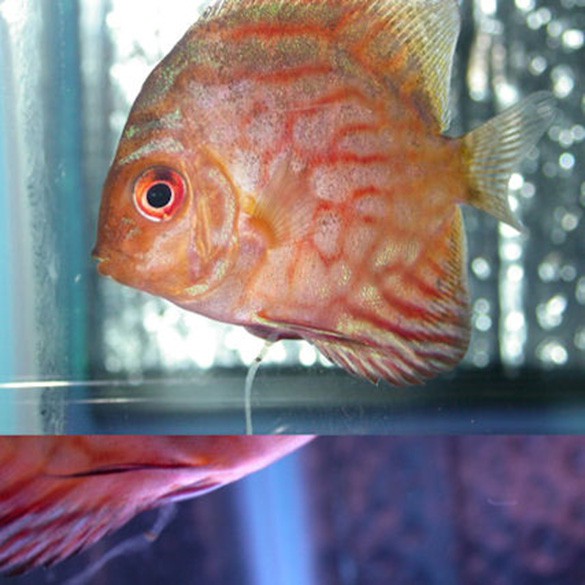
- سیاه شدن پوست
- بی رنگی آبشش
- آسیت
- اتساع محوطه بطنی
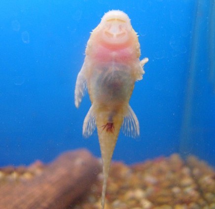
- اگزوفتالمی
- خونریزی های سر سوزنی بر روی سطح خارج و محوطه بطنی
- علام عصبی (شنای چرخشی، flashing، شنای ناگهانی)
- ناهنجاری های نخاعی به صورت لردوزیس و اسکولیوزیس
دامنه میزبانی
آزاد ماهیان
قزل آلای رنگین کمان
آزاد ماهیان سیاه
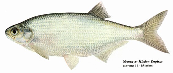
آزادماهی چشم قرمز
سگ ماهی آزاد
آماگو
آزاد ماهی ژاپنی
آزاد ماهی نقره
آزاد ماهی اطلس
سایر آزاد ماهیان و برخی غیرآزاد ماهیان گاهی می توانند به بیماری مبتلا شوند
علایم کالبدگشایی
کم خونی
عدم وجود غذا در روده ها
کبد، کلیه و طحال رنگ پریده
مایعات آسیتی و خونریزی های سرسوزنی در اعضای مختلف محوطه بطنی
آسیب شناسی
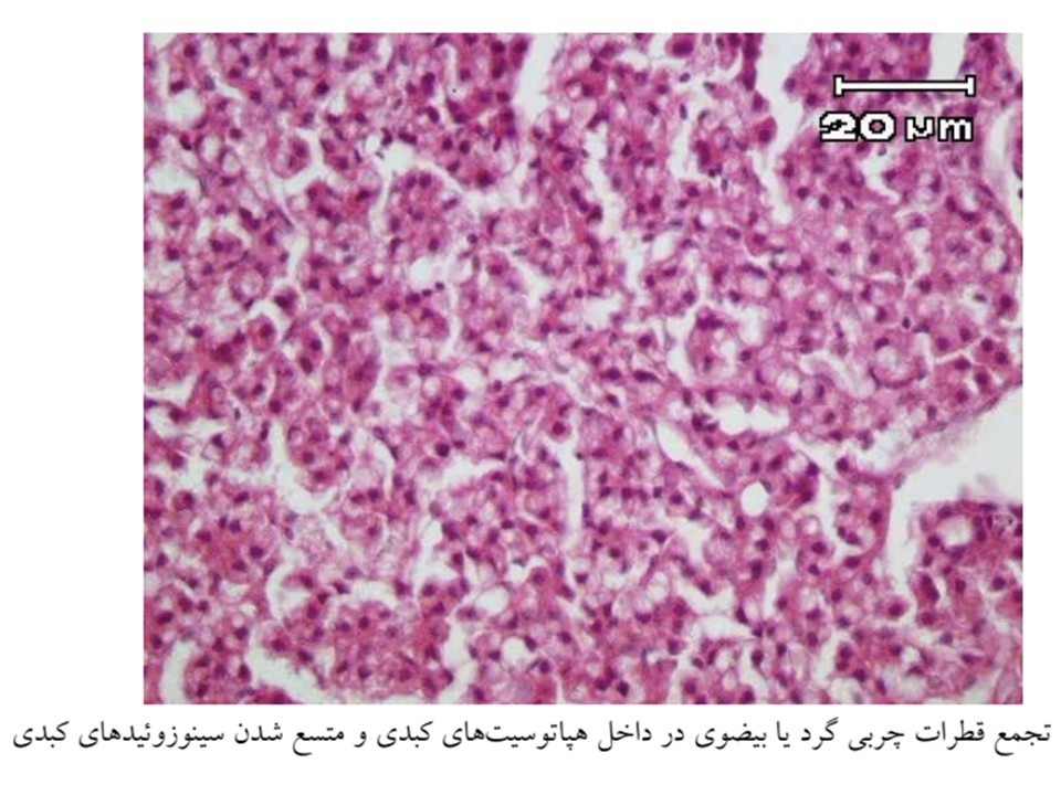
نکروز دژنراتیو بافت های خونساز، کلیه، طحال، کبد، پانکراس و دستگاه گوارش
نکروز سلول های گرانولار ائوزینوفیلیک در دیواره روده ها (وجه تشخیص ازVHS)
گنجیدگی های نکروبیوتیک و ماکروفاژهای کف آلود
لکوپنی، دژنرسانس گلبول های سفید و ترومبوسیت ها و تعداد زیادی بقایای سلولی
کاهش بیلی روبین، بیکربنات، کلسیم، کلریدها و فسفر در موارد حاد بیماری
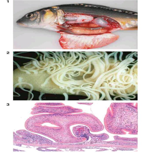
Salmonid with IHN. Note the characteristic, thick trailing fecal cast
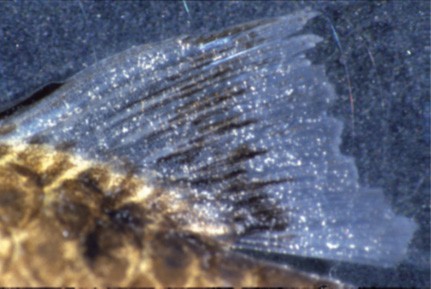
Exopthalmia with haemorrhage in rainbow trout fry with infectious haematopoietic necrosis
Histological section showing acute necrosis of eosinophilic granular cells (arrows) of the intestinal submucosa. H&E
Focal necrosis of kidney interstitium of rainbow trout with
infectious haematopoietic necrosis. Bar = 100 μm
Pyknotic and karryorrhectic nuclei in haematopoietic tissue in head kidney in sockeye salmon fry with infectious haematopoietic necrosis. Note pale eosinophilic adrenal cells with large nuclei
Liver of sockeye salmon fry with infectious haematopoietic necrosis. Hepatic necrosis with strong positive immunohistochemical reaction. Bar = 100 μm
Intestine of sockeye salmon fry with infectious haematopoietic necrosis. Strong positive immunohistochemistry reaction in stratum proprium. Bar = 50 μm
Histological section showing acute necrosis of kidney hematopoietic tissue (H) caused by IHN. note the lack of damage to renal excretory tissue. H&D
smear with necrobiotic body (arrows) caused by IHN. Giemsa
IHN in Atlantic salmon, Salmo salar, section of the heart. A necrotic thrombus is evident with foci of erythrocytes indicating haemorrhage. ×80.
IHNV immunohistochemistry (IHC) in different tissues of steelhead fry by experimental infection. Pre-epizootic stages (0 –5 days) contrast with epizootic stages (6 –14 days). (A) Gill, pre-epizootic stage, with IHC-positive lamellar and fi lament epithelial cells (ep, arrow). (B) Gill, epizootic stage, with IHC -positive lamellar and fi lament endothelial cells (en, arrow). (C) Anterior kidney, pre -epizootic stage, with IHC -positive tubule (t, arrow) and negative haematopoietic cells. (D) Anterior kidney, epizootic stage with IHC -positive haematopoietic cells (h, arrow). (E) Pyloric caeca, pre -epizootic stage, with IHC -positive columnar epithelial cells (ep, arrow). (F) Intestine, epizootic stage, with negative columnar cells and IHC -positive lamina propria (lp, arrow). (G) Brain glial cells, epizootic stage, with IHC -positive glial cells (g, arrow) in the granular layer of the optic lobe. (H) Eye, epizootic stage, with IHC -positive retina (r, arrow). a –f ×40. g ×40. h ×10mag