بیماری های ماهی ناشی از سخت پوستان
بیماری های ناشی از سخت پوستان
ملیکا باعث
جهت مشاهده متن کامل مقاله اینجا کلیک کنید
Argulus Infestation: Fish Louse
branchiuran crustaceans(most important is Argulus).
uncommon in freshwater aquarium fish but may occur if wild or pond raised fish are introduced into the tank and infest the skin and fins of numerous fish worldwide.
common on goldfish and koi and can be prevalent on many wild freshwater fish (cyprinids, centrarchids, salmonids).
Common marine hosts include the mummichog, gulf killifish and sheepshead minnow.
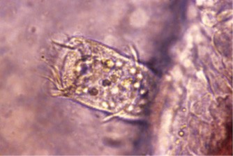
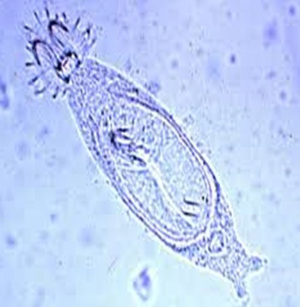
Whole mount of Argulus coregoni from the skin of a wild grayling.
The structure of an argulid (adult male).
Many fish lice have a wide host range:
Argulus foliaceus:
trout fisheries
increasing losses
reducing the appetite of infected fish
making them less likely to react to bait
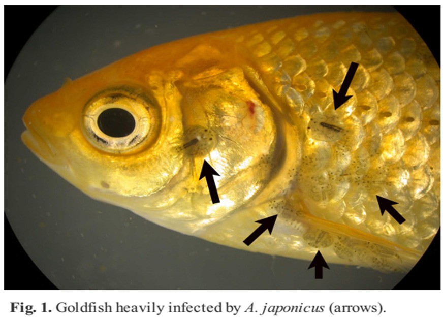
Argulus japanicus: originally from the Orient, has been spread worldwide on goldfish.
Argulus coregoni: is the largest of the three species and measures up to 13 mm in length.
Pathogenesis
Fish lice feed by inserting a pre-oral sting (stylet) into the host and sucking body fluids.
Fish can display violent erratic swimming or other behavioral abnormalities because of the irritation caused by the stylet.
Fish are damaged by the repeated piercing of the skin by the stylet, which injects toxic enzymes, causing irritation.
This irritation may cause focal hemorrhage or hyperpigmentation. Fish may be anemic.
Argulus can also mechanically transmit bacterial (Pseudomonas spp. and Aeromonas Spp) or viral pathogens (SVC).
Fish lice can be intermediate hosts for several fish-parasitic nematodes, including members of the families Anguillicolidae, Skrjabillanidae, and Dracunculoidea.
One or two parasites usually cause no clinical signs in large fish, but fish lice have a high reproductive rate, often resulting in rapid escalation of infestations.
Clinical Signs
- jump repeatedly, scrub against surfaces, stop feeding and appear dark.
- Histologically, necrosis and epidermal proliferation occurs around entry wounds and haemorrhage with a severe inflammatory response involving lymphocytic infiltration, typically follow.
- peak abundance typically in summer and fall.
- The entire life cycle is typically 30 days or more. Eggs laid on vegetation or other objects usually hatch into juveniles within 10–50 days.
- Juveniles (1– 3mm) look like adults without suckers; they must find a host within 2–3 days or will die.
- Adults can also survive without a host for several days.
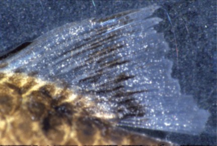
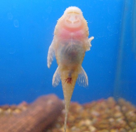
Heavily infested Koi. Note readily visible oval parasites in throat (ventral) area of head, as well as others scattered throughout the body.
Diagnosis
- History:
Pruritus/itching ; red sores; wild-caught or pond-raised fish.
- Physical Examination:
Focal red lesions on skin; focal color change (especially darkening) on skin.
- Method of Diagnosis:
Wet mount of skin or buccal cavity with parasite.
- Fish lice frequently move on a host and may be seen swimming when they are in an aquarium. They often remain attached when the host is removed from the water. Fish lice look like a moving fish scale.
Treatment
The life cycle of parasite varies but typically is about 2 months.
it is useful to rid tanks of egg contamination by using disinfectant.
allowing the tanks to dry thoroughly for several days.
Organophosphate prolonged immersion.
Formalin bath.
Potassium permanganate bath.
Enamectin oral.
Individual parasites can be removed from fish by using forceps, but this does not eliminate parasites in the environment and smaller individuals might be missed.
Lernaeids (Anchor Worms)
Anchor worms may be introduced into an aquarium from wild or pond-raised fish. Goldfish, koi, or wild native fish are most commonly affected.
Lernaea and related genera infect freshwater fish. In temperate climates, Lernaea is most likely to be seen in summer, when reproduction usually occurs.
After several nonparasitic stages, the terminal copepodid stage attaches to a fish, mates, and the male dies. The female then penetrates under the skin of the fish and differentiates into an adult.
Clinical signs
- Single lernaeid parasites are usually not life- threatening, unless they are infecting a small fish or when they penetrate near vital organs.
- Heavy infections can lead to debilitation and secondary bacterial or water mold infection.
- History/Clinical signs:
Hemorrhage at the site of attachment.
considerable hyperplasia or fibrosis may develop at the attachment site.
skin sores.
- Physical Examination:
Various-sized (barely visible to ~ 25mm) copepods attached to gill arches, oral cavity, or skin; erosion and/or ulceration; red areas on skin, may be raised up to 5mm in height.
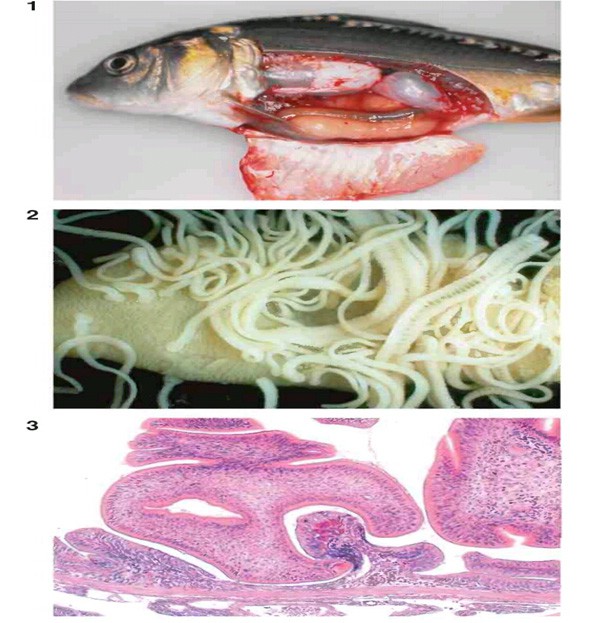
Anchor worm (Lernaea cruciata) infection of a largemouth bass. The head of the parasite is embedded under the skin while the body (P) with egg sacs protrudes. Note the hemorrhage (arrow) where the parasite enters the fish. PF = pectoral fin.
Lernaea polymorpha parasites on a bighead carp.
Method of Diagnosis:
- Wet mount of gills, skin, or mouth with parasite.
- Histopathology of gills, skin, or mouth with parasite.
- Diagnosis of copepod infection is based on identification of typical parasitic life stages on fish:
- Large, mature females are often pathognomonic.
- Small immature stages, such as copepodids or even immature adults, may not be grossly visible, so microscopic examination of skin scrapings is advisable.
- Histopathology may also be used to identify permanently attached forms.
Treatment
- Organophosphate prolonged immersion(should be repeated every 7 days for 28 days)
- Difluorobenzuron prolonged immersion.
- Salt prolonged immersion (freshwater copepods, 20– 40mg/L).
- removing individual parasites with forceps. Note that larval stages may still remain on the fish or in the water; thus, fish must still be treated and then placed in uncontaminated water. Treating with potassium permanganate after removal of adults can be curative.
Ergasilids
they are also modified for parasitism as indicated by the presence of antennae modified for grasping the host and a large trunk for reproductive products.
Ergasilids are usually ∼2mm long, with a conical, segmented body. Only the female of the species are parasitic on fish, all other stages being free living.
Most infest the gills (rarely the skin) of freshwater fish. Some have a wide host range (Neoergasilus japanicus and Ergasilus sieboldi on temperate freshwater fish; E. labracis on temperate marine fish).
Rarely causing epidemics in cultured fish, they are often incidental findings on wild or pond-raised fish in summer and probably cause few problems in small numbers.
Clinical signs:
Severe gill damage is caused by the feeding activity of the copepods and very heavy infestations can be lethal. The gills are anaemic and secondary bacterial infections can result
Adult females of some species can move from host to host. Several ergasilids have been inadvertently spread across continents by movement of infested fish
Method of Diagnosis:
- Wet mount of gills or skin with parasite
- Histopathology of gills or skin with parasite
Treatment:
organophosphates. For example, Neguvon® (0.25mg/L) controlled Ergasilus labracis on Atlantic salmon parr. There is no evidence of resistance in fish recovering from natural infestations
Ergasilus female removed from rainbow smelt gill.
(a) Adult female ergasilid.
(b) Adult female lernaeid
male Ergasilus.
Adult female Ergasilus on gill of rainbow smelt
Severe gill maggot (Ergasilus) infestation of a striped bass.
The parasites are attached to the primary lamellae of the gills by their modified antennae. Note that the egg sacs (E) vary from white to grey, depending on the developmental stage of the larvae in the egg sacs.